

Medical imaging has developed considerably over time, enabling doctors to identify illnesses with better precision during detection and treatment while monitoring patients’ conditions. The PET CT scan stands out today as a prominent diagnostic instrument for detecting many medical conditions among various modern imaging technologies. This combined functional and anatomical imaging capability of PET together with CT yields detailed pictures of the entire body.
Healthcare professionals recognize that PET CT scans have become essential resources for modern clinical diagnosis practices regardless of cancer brain disease or heart disease cases. This article examines the ten most frequent medical conditions that health professionals identify through PET CT scanning procedures.
1. Cancer Diagnosis and Staging
PET CT scans find widespread use in cancer diagnosis because they help doctors stage cancer from its beginning to its most advanced stage. Through the use of a PET scan for cancer, abnormal cell detection is possible alongside cancer metastasis monitoring and ongoing treatment assessment. PET CT provides superior cancer diagnosis because it finds early tumours by uncovering regions of elevated metabolic activity that typically signal growing tumours. Different types of cancers use this imaging technique for diagnosis purposes.
Types of Cancers
- Lung cancer
- Breast cancer
- Colorectal cancer
- Lymphoma
Scheduled PET CT scans support cancer patients in their treatment through tumour response evaluation which guides treatment adjustment.
2. Brain Disorders and Neurological Conditions
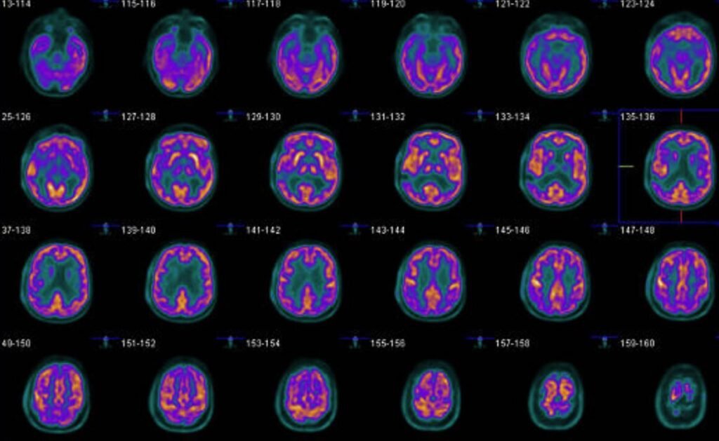
Medical imaging plays a vital role in detecting complex brain disorders because the brain remains one of the most intricate organs within the human body. PET CT scanners enable vital neurological condition diagnosis through their capability to examine brain processes, blood circulation patterns and metabolic activity.
- Alzheimer’s disease
- Epilepsy
- Parkinson’s disease
The clear visual display of plaque accumulation in brain areas provides physicians with the ability to perform early interventions for Alzheimer’s disease patients.
3. Cardiovascular Diseases
Through PET CT scanning physicians can diagnose cardiovascular conditions while using these scans to perform accurate evaluations of heart diseases. The assessment through a PET CT scan helps doctors measure heart muscle blood flow patterns while locating areas of reduced functioning and quantifying heart damage following a heart attack. Conditions commonly evaluated include:
- Coronary Artery Disease (CAD)
- Cardiomyopathy
- Congestive Heart Failure
The precise demonstration of heart functional imaging through this technology enabled successful proactive medical treatment which saved numerous lives.
4. Lung Diseases
Health issues of the lungs encompass both milder infections as well as critical cancers. The medical field uses PET scan for cancer as a diagnostic tool to inspect lung nodules along with their malignancies or to establish lung cancer extents. PET CT scans provide vital information about lung disease to healthcare providers who work with patients who have emphysema or COPD (Chronic Obstructive Pulmonary Disease).
Pet CT Scan in Bangalore operates trustworthy diagnostic facilities that utilize leading-edge imaging technologies to perform such diagnostic tests.
5. Infections and Inflammatory Conditions
PET CT technology extends beyond its use for cancer diagnosis and treatment of chronic diseases. These diagnostic tools prove to be useful for identifying infections as well as inflammatory conditions in conditions where other diagnostic methods are not effective. A PET CT scan enables doctors to recognize specific conditions through the following examples:
- Osteomyelitis (infection of the bone)
- Infected prosthetic devices
- Fever of unknown origin (FUO)
The scan shows a more accurate depiction of internal body processes by detecting places infected or affected by inflammation.
6. Thyroid Cancer and Disorders
PET CT scans successfully identify thyroid cancer which represents a widespread medical condition among patients. PET CT offers highly effective detection of thyroid cancer remnants that standard imaging such as ultrasound and X-ray examinations fail to identify. The scan provides information about thyroid nodules by determining their risk level between benign and malignant conditions.
7. Esophageal and Stomach Cancers
The early signs of esophageal and stomach cancer are often confused with other related digestive conditions thus making these cancers challenging to detect at an early stage. A PET scan for cancer helps resolve detection challenges because it reveals signs of metabolic activity in these types of cancers. PET CT scans help doctors assess the stage of development of cancers while enabling them to determine their spread through other body parts. Treatment teams create the best treatment plans through exact disease identification which helps prevent unnecessary invasive methods.
8. Lymphoma
PET CT scans find widespread application in lymphoma diagnosis since this type of cancer develops in the lymphatic system. PET CT scans prove useful for diagnostic evaluation, staging assessment and therapeutic evaluations. PET CT scans show whether cancer tumours have invaded lymph nodes situated beyond the original tumour site. PET CT offers detailed lymphatic system analysis to enable medical professionals to identify early diseases and develop proper therapeutic approaches.
9. Bone Metastases and Bone Disorders
Diagnostic assessment of bone metastases (cancer spread to bones) together with bone infections remains difficult through conventional imaging tools. PET CT scanning technology reveals necessary bone information since it shows details of both structural elements and metabolic activities within scanned areas.
10. Full Body Screening for Preventive Health
PET CT scans originally served diagnostic functions but medical practitioners now use them to assess patient health through preventive examinations. Most patients seeking full body checkup in Bangalore and other urban areas now receive This scan during their examinations to screen multiple organs and systems.
The early identification of cancers together with cardiovascular diseases and disorders happens before symptom manifestation permitting healthcare providers to start interventions that lead to positive results.
Final Thoughts
The PET CT scan has established itself as a primary diagnostic and monitoring tool for numerous medical conditions because of continuous medical imaging progress. PET CT combines functional imaging capabilities which transforms it into vital equipment for the identification of several health conditions including cancers and brain disorders alongside heart problems.
The evolution of diagnosing techniques in modern medicine began through the implementation of this technological method which enables accurate detection of conditions and effective treatment evaluation.
The pet CT scan in Bangalore as well as other reputable diagnostic centers located in Bangalore operates with accredited professionals who maintain high-quality imaging capabilities. Patients who want preventive care should consider undergoing full body checkup in Bangalore examinations which detect early problems so future health remains strong.
PET CT scans have delivered remarkable progress that makes medical diagnosis and illness treatment both faster and more accurate for the medical profession.
Koshikaa stands as a trusted healthcare provider that delivers extensive full body checkup in Bangalore supported by state-of-the-art diagnostic services to detect health problems at an early stage.